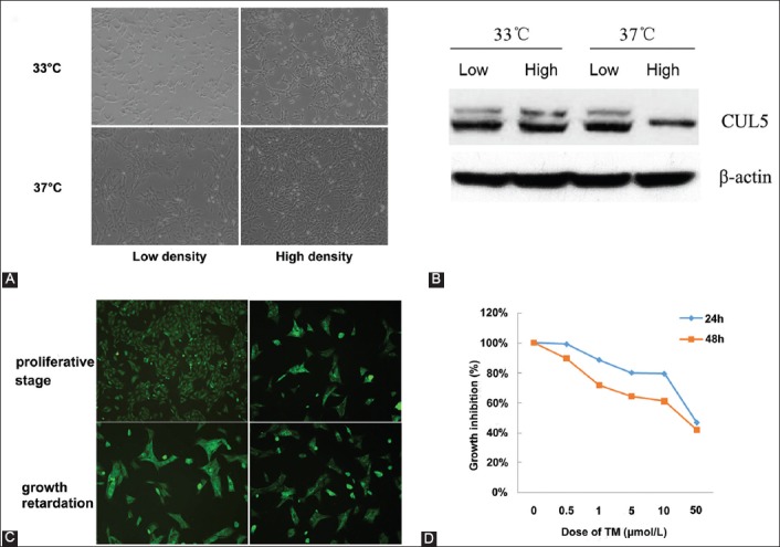
FIGURE 1.

The tunicamycin (TM)-induced MPC5 model construction. A: The MPC5 cell proliferation was increased by a high dose of TM at 33°C, while the cell differentiation was induced by a high dose of TM at 37°C; B: Western blotting showed that the expression of Cullin-5, a cell proliferation indicator, was decreased by a high dose of TM compared to low dose TM treatment in the MPC5 cells; C: The immunofluorescence assay showed that the MPC5 model cells were successfully constructed; D: The cell viability assay revealed that the optimal concentration of TM for MPC5 cells was around 1-10 µmol/l.
