
Figure 2. Range of Choriocapillaris (CC and Retinal Pigment Epithelium Alterations in Patients and Carriers in Coregistered Images.
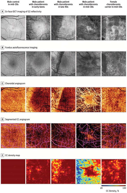
A, En face optical coherence tomography (OCT) of ellipsoid zone (EZ) reflectivity demonstrates progressive EZ loss. B, Fundus autofluorescence imaging demonstrates relative preservation of retinal pigment epithelium autofluorescence corresponding to regions of intact EZ. Retinal pigment epithelium loss is more extensive than EZ loss in nearly all eyes. C, Choroidal angiogram demonstrates increasing degrees of CC atrophy with exposure of underlying choroidal vessels. D, Segmented CC angiogram demonstrates that CC density is subnormal in affected eyes throughout tine imaged field but is worse underlying regions of EZ and retinal pigment epithelium loss. Projection artifact from large inner retinal vessels is indicated in purple. E, Mapping of CC density highlights areas of CC loss.