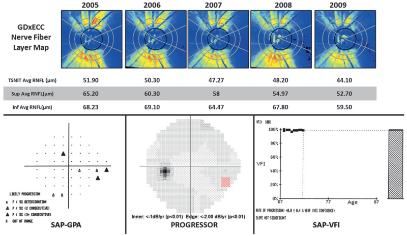
Figure 2.
GDx with enhanced corneal compensation (GDxECC) retardation maps (upper row) showing progressive loss of the superior retinal nerve fibre layer (RNFL) in a 74-year-old female with open-angle glaucoma over 4 years of follow-up. Standard automated perimetry Guided Progression Analysis based on EMGT criteria (SAP-GPA, bottom left) and Progressor (bottom centre) demonstrates progressive inferior nasal visual-field loss. The visual-field index (VFI, bottom right) slope shows no significant change over time. TSNIT, temporal superior nasal inferior temporal.

