
Figure 4.
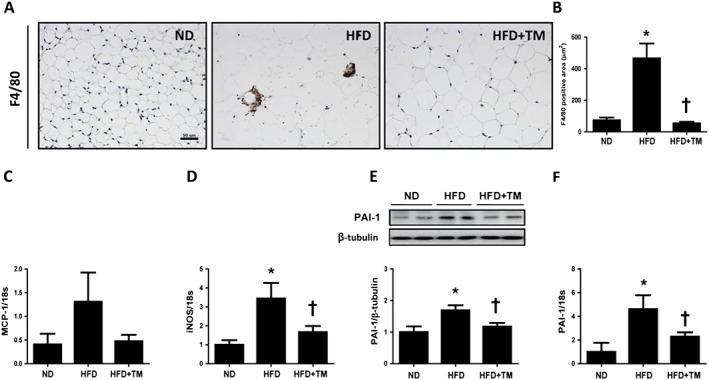
TM5441 prevents HFD‐induced inflammation in the epididymal WAT. (A and B) Representative immunohistochemical staining of F4/80 (1:200) and quantification of F4/80‐positive area in epididymal WAT. Magnification, 100×; scale bar, 100 μm. (C and D) MCP‐1 and iNOS mRNA levels were determined by quantitative reverse transcription PCR (qRT‐PCR). (E and F) PAI‐1 protein and mRNA expression in the epididymal WAT were determined by western blot analysis and qRT‐PCR. The data are shown as the mean ± SEM of seven mice. * P < 0.05 versus ND, † P < 0.05 versus HFD.