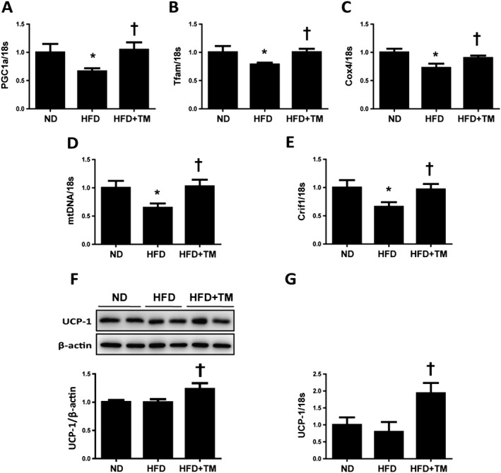
Figure 5.

TM5441 restores HFD‐induced mitochondrial dysfunction in the epididymal WAT. (A–E) Total RNA was isolated from the epididymal WATs and subjected to quantitative reverse transcription PCR analysis to determine the expression of (A) PGC1α, (B) Tfam, (C) Cox4, (D) mtDNA, (E) Crif1 and (G) UCP‐1. Expression was normalized against 18S rRNA levels. (F) UCP‐1 protein expression level was detected by western blot analysis, where β‐actin was used as an internal control. The data are shown as the mean ± SEM of seven mice. * P < 0.05 versus ND, † P < 0.05 versus HFD.
