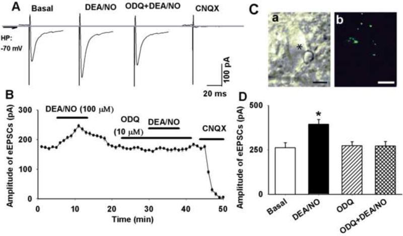
Figure 1. DEA/NO increased evoked EPSCs in second-order NTS neuron.

A: Raw tracings showing TS-eEPSCs in a labeled NTS neuron during control and application of 100 μM DEA/NO, DEA/NO plus ODQ (10 μM), and CNQX (20 μM). The traces are averages of 10 consecutive responses. B: ODQ abolished DEA/NO-induced increase in eEPSCs in a labeled NTS neuron. C: an NTS neuron (indicated by *) viewed with infrared light and differential interference contrast optics (a) and the attached DiA-labeled baroreceptor afferents terminals (b, green color). D: summary data showing that DEA/NO increased amplitude of TS-evoked EPSCs in second-order NTS neuron, an effect was abolished by ODQ. Data were presented as means ± SEM. * P < 0.05 compared with control value. Scale bars in C a and b: 10 μm.
