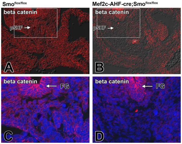
Fig. 6.
Loss of Smoothened from the SHF results in a reduction of β-catenin expression in the pSHF. A,C: The normal expression of β-catenin in the venous pole of a ED10.5 Smofl/fl control embryo (C is an enlargement of the boxed area in A). B,D: The reduced expression of β-catenin expression in a SHF-Smofl/fl littermate. Note that the β-catenin staining in the foregut endoderm is not affected by conditional deletion of Smo from the SHF. Blue staining in C and D is DAPI nuclear stain. FG, foregut; pSHF, posterior second heart field

