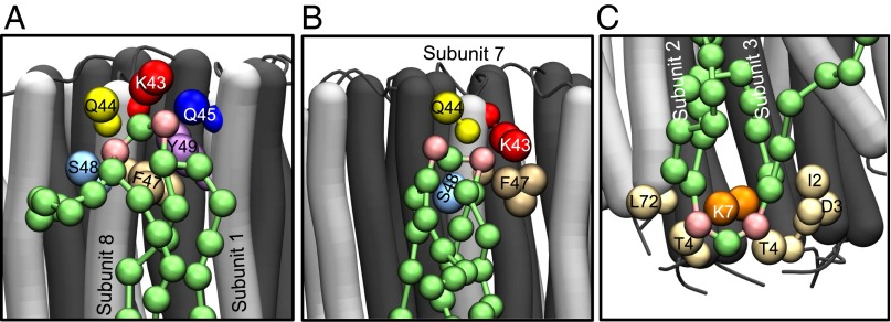
Fig. 3.
Modes of binding of cardiolipin to trimethylated c8-rings. The N- and C-terminal α-helices of c-subunits are dark and light gray, respectively. Cardiolipin molecules are green, with pink phosphate groups. The large colored spheres represent the coarse grain beads for specific amino acids, as indicated, lying within 0.7 nm of the phosphate beads of cardiolipin. (A and B) Head-group region of cardiolipin in the inner leaflet of the membrane bound, respectively, to two adjacent c-subunits (subunits 8 and 1) and to a single c-subunit (the relative frequency of these two binding modes is shown in Fig. S12). (C) A cardiolipin molecule in the outer leaflet of the membrane bound to a single c-subunit.

