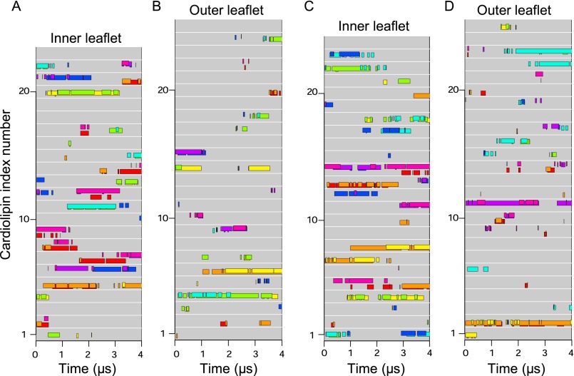
Fig. S5.
Individual cardiolipins bound to distinct c-subunits in inner and outer leaflets during both simulations of the demethylated c8-ring. (A and B) First simulation. (C and D) Second simulation. Data are shown for cardiolipin molecules in the inner (A and C) and outer (B and D) leaflets of the membrane. Each gray line represents a cardiolipin, and colored bars along the line indicate when a cardiolipin phosphate bound to a c-subunit, as indicated by bar color. The c-subunits s1–s8 are colored as follows: s1, red; s2, orange; s3, yellow; s4, green; s5, cyan; s6, blue; s7, purple; s8, magenta.

