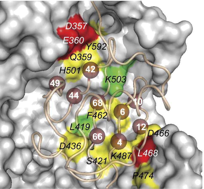
Fig. 3.
Alanine-scanning of USP2 site 2. The structure of USP2 bound to Ubv.2.1 (PDB ID code 3V6C) is shown. USP2 is shown as a surface colored according to the fold reduction in catalytic efficiency relative to WT caused by alanine substitutions at the labeled positions, as follows: green < 2; 2 ≤ yellow < 10; red > 10; gray, unscanned. The Ubv02.1 main chain is shown as a tube, and residues in the core functional epitope are shown as numbered spheres.

