Fig. 5.
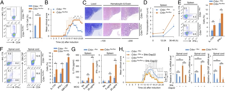
Exacerbated and sustained EAE symptoms in T-cell–specific Crbnflox/flox;Cd4-Cre mice. (A) Flow cytometry of IL-17A–producing CD4+ T cells among the in vitro-differentiated Th17 cell population within CD4+ T cells from Crbnflox/flox;Cd4-Cre (flox/flox) or Crbn+/flox;Cd4-Cre (+/flox) mice. (B) Clinical scores for EAE mice (n = 7 per group). (C) Spinal cord sections. (D) Proliferation of splenocytes in response to MOG35–55, was measured by [3H]thymidine incorporation, at the indicated time points for 12 h. (E) Percentage of CD4+IL-17A+ and CD4+IFN-γ+ T cells in the spleens of mice after EAE induction (n = 3 per group). (F) Percentages of CD4+IL-17A+, CD4+IFN-γ+, and CD4+GM-CSF+ T cells in the spinal cords of mice after EAE induction (n = 3 per group). (G) Concentration of cytokines in the supernatant from splenocyte cultures stimulated with MOG35–55 peptide (n = 3 per group). (H) Clinical scores for EAE mice (n = 8 per group) with or without Shk-Dap22 treatment. (I) Relative mRNA levels of IL-17A, IFN-γ, and GM-CSF in isolated spinal cord mononuclear cells (n = 3 per group). Data are representative of five (A), three (B–G), or two (H and I) independent experiments. Results are expressed as mean ± SD. *P < 0.05; **P < 0.01, unpaired two-tailed Student’s t test.

