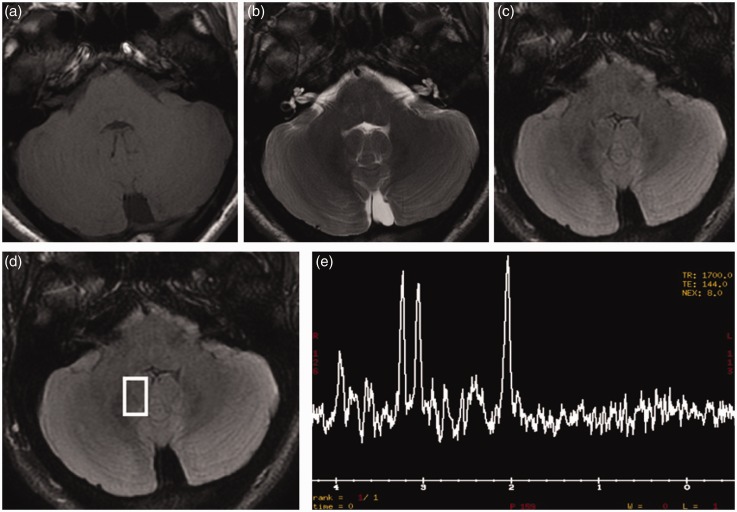
Figure 5.
Brain magnetic resonance imaging (MRI) obtained six months after diagnosis. (a) T1, (b) T2, (c) fluid-attenuated inversion recovery (FLAIR) images show disappearance of the cerebellar lesions. Spectroscopy (d) and (e), acquired within the affected areas, showed normalisation of the spectrum.

