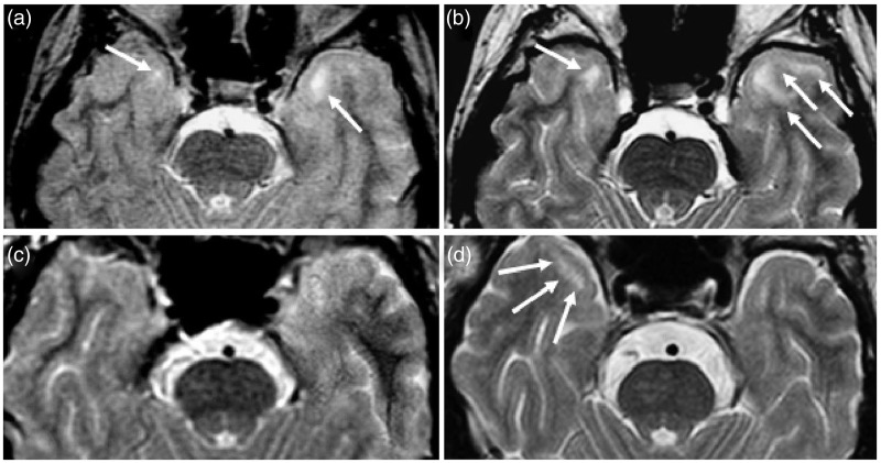
Figure 3.
Axial T2-weighted images showing the enlargement of pre-existent temporopolar white matter lesions (WMLs) (a) and (b) in patient 3 (Table 2) and the development of a temporopolar WML (c) and (d) in patient 5 (Table 2) during follow-up: (a) and (c) (baseline), (b) (19 years later) and (d) (16 years later).

