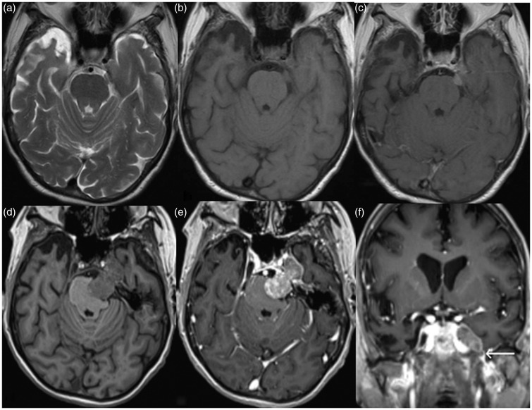
Figure 1.
Axial T2, pre and post gadolinium T1-weighted magnetic resonance (MR) images (a), (b), and (c) demonstrate a small T2 hypointense homogeneously enhancing dural-based mass along the left petrous apex extending into the left prepontine cistern, and a diagnosis of meningioma was considered. Four months later, the symptoms progressed, and follow-up axial pre and post gadolinium T1-weighted MR images ((d) and (e)) reveal a large heterogeneously enhancing mass along the expected course of left trigeminal nerve resulting in effacement of the left ventral pons with extension into the left cavernous sinus and asymmetric effacement of left Meckel’s cave. Based on the imaging characteristics and minimal inferior extension into the foramen ovale (white arrow) on corresponding coronal post gadolinium T1-weighted image (f), a trigeminal schwannoma was suspected. A carcinoid tumor metastatic to meningioma was found on histology.

