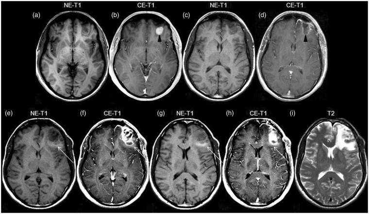
Figure 3.
Initial, preoperative MR showing a left frontal lesion, hypointense on T1WI (a) and presenting contrast enhancement (arrowhead in b). Follow-up MR, three months after surgery ((c), (d)), shows linear enhancement in the periphery of the excised area (arrowhead in (d)) and lack of edema. Next MR, six months after surgery ((e)–(i)), demonstrates recurrence with significant contrast enhancement ((f), (h)). Notably, hyperintense edema on T1WI is now present (arrow in (e)–(h)). T2WI demonstrates greater extension of the edema (i). T1WI: T1-weighted imaging; NE-T1: nonenhanced T1-weighted imaging; CE-T1 contrast-enhanced T1-weighted imaging; MR: magnetic resonance.

