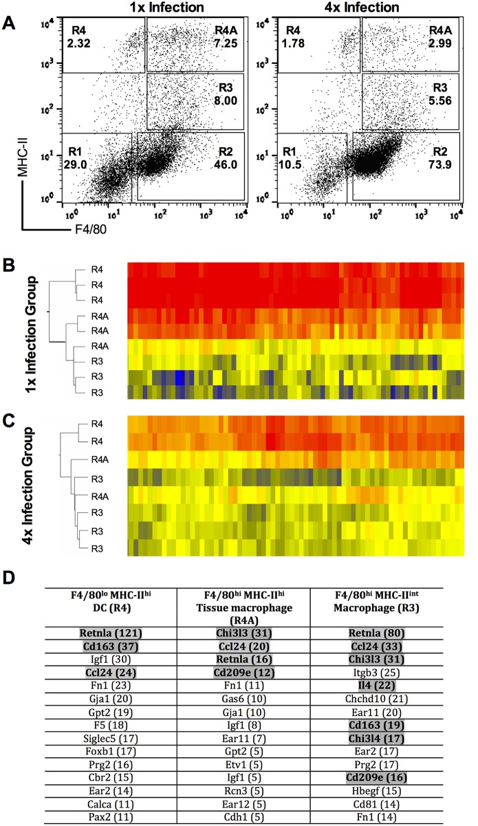
Fig 1. Genes related to alternative activation cell phenotype are up-regulated in dermal exudate cells (DEC) populations after repeated infection.
A. Representative flow cytometry dot plots for DEC showing the distribution of cells based upon their expression of F4/80 and MHC-II. Cells were gated into R4, R4A and R3 populations recovered from 1x (left) and 4x (right) infected mice. Values in bold are the percent positive cells found within each gate expressed as a proportion of the total DEC recovered. Cells were sorted by fluorescent activated cell sorting (FACS), and RNA from the sorted R4, R4A and R3 cell populations was applied to microarray analysis. Heat maps showing the clustering of genes within each sorted DEC population for three biological replicates recovered after B. a single (1x) infection, and C. repeated (4x) infections. D. Identity of the top 15 up-regulated genes in each population after 4x compared to 1x infection. Number in brackets represents the fold up-regulation of that gene in the sorted cell population from 4x infected mice. Grey shaded genes represent those that are associated with alternative activation.

