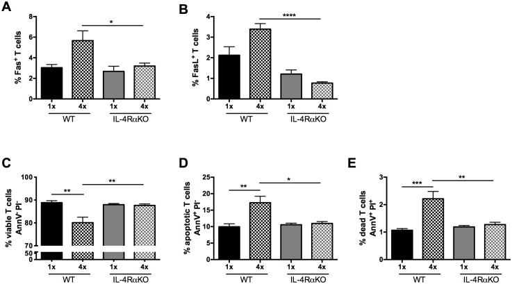
Fig 5. Expression of Fas/FasL and viability of CD4+ T cells in the sdLN is enhanced in the absence of IL-4Rα.
Percentage of CD4+ T cells that are A. Fas+ and B. FasL+ in 1x and 4x infected WT compared to IL-4RαKO mice. C-E Percentage of viable (AnnV-PI-), apoptotic (AnnV+PI-) and dead (AnnV+PI+) CD4+ T cells in the sdLN of 1x and 4x infected mice as determined by an AnnexinV assay. n = 3–4 mice per group; n.s. denotes ‘not significant’ p>0.05; * = p<0.05; ** = p<0.01; **** = p<0.0001 as determined by ANOVA and Tukey’s post-test analysis.

