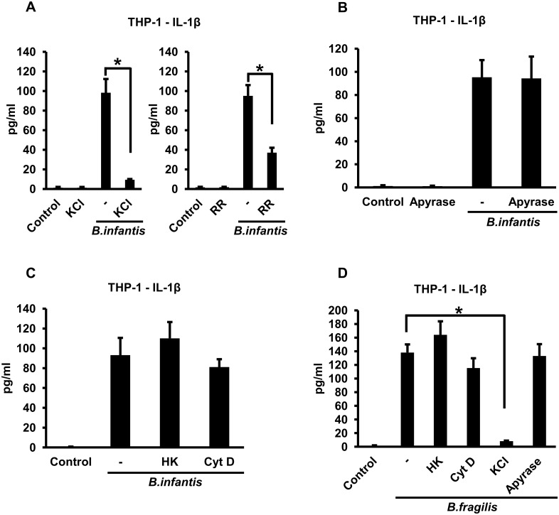
Fig 5. B. infantis- and B. fragilis-induced IL-1ß secretion by THP-1 macrophages is dependent on potassium efflux but does not require bacterial viability or phagocytosis.
A. THP-1 macrophages were infected with B. infantis for 1 hour in the presence or absence of 50 mM potassium chloride (KCl) or 2 μM ruthenium red (RR), as indicated. The cells were washed and incubated in fresh medium, with potassium chloride or ruthenium red added back as appropriate. Supernatants were collected at the end of 4 hours and IL-1ß concentrations determined by ELISA. *p < 0.0001, n = 6 per experimental condition. B. THP-1 macrophages were infected with B. infantis for 1 hour. The cells were washed and incubated in fresh medium, with 5 units/ml of apyrase added as indicated. Supernatants were collected after 4 hours and IL-1ß concentrations determined by ELISA, n = 6 per experimental condition. C. THP-1 macrophages were infected with equivalent numbers of live or heat-killed (HK) B. infantis for 1 hour, in the presence of 5 μM cytochalasin D (CytD) where indicated. The cells were washed and incubated in fresh medium, with cytochalasin D added back as appropriate. Supernatants were collected after 4 hours and IL-1ß concentrations determined by ELISA, n = 6 per experimental condition. D. THP-1 macrophages were infected with equivalent numbers of live or heat-killed (HK) B. fragilis for 1 hour, in the presence of 5 μM cytochalasin D (CytD) or 50 mM potassium chloride (KCl) where indicated. The cells were washed and incubated in fresh medium, with cytochalasin D, potassium chloride or 5 units/ml of apyrase added back as appropriate. Supernatants were collected after 4 hours and IL-1ß concentrations determined by ELISA. *p < 0.0001, n = 6 per experimental condition.

