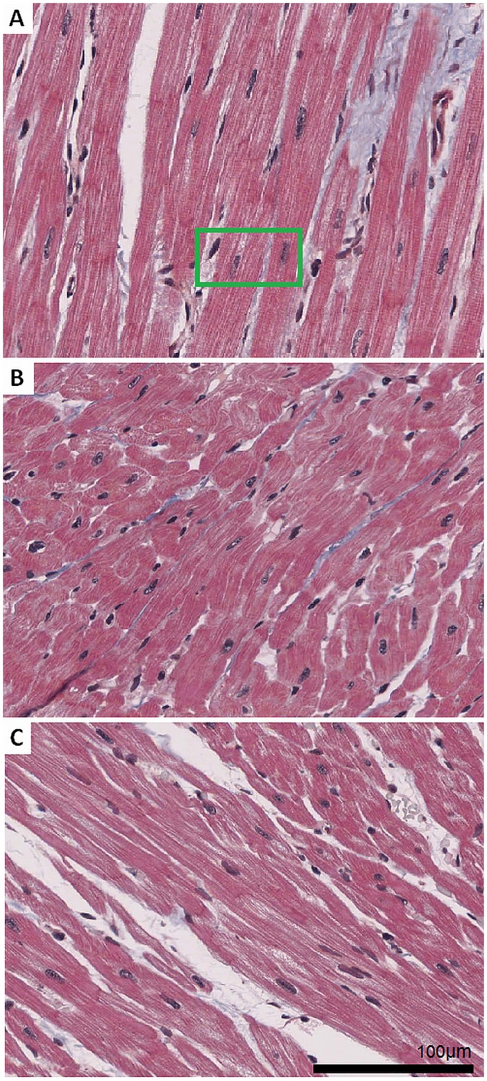
Fig 1. Sample images of cardiac tissue.

Heart tissue slices were stained using the Trichrome (Gieson) protocol and scanned with a high resolution scanner. Sample images are cut-outs with a size of 1024 x 768 pixels. (A) Micrograph image section with fibres predominately oriented parallel to the image plane. Details of the green region of interest (ROI) are depicted in Fig 2. (B) Tissue with myocytes oriented rather oblique and perpendicular to the image plane. (C) Region with cells mixed in orientation from parallel to perpendicular to the image plane.
