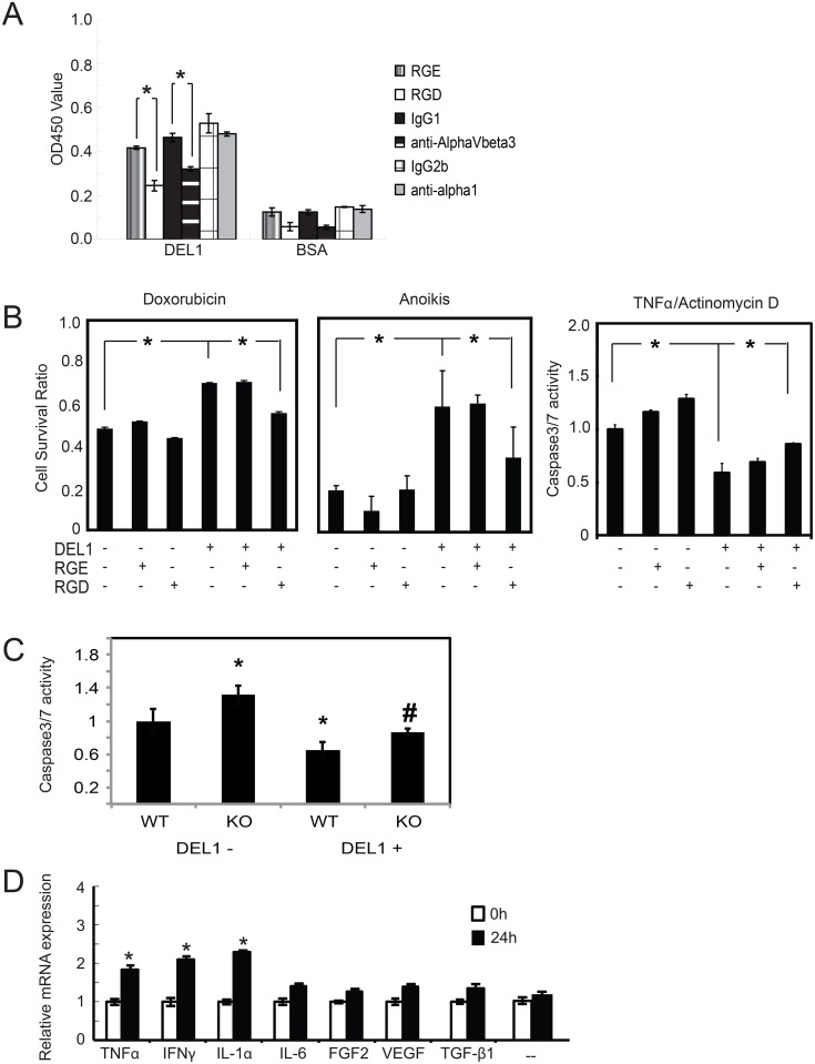
Fig 3. DEL1 effect on apoptosis and induction.
(A) NHACs were pre-treated with the peptides or antibodies indicated and placed in plates coated with either BSA or DEL1. Cells attached after 6 hrs were determined by WST-8 assay. *p<0.05 between indicated values. (B) NHACs cultured with DEL1 have increased survival after pro-apoptotic stimuli that were inhibited by RGD, not RGE, peptides. For caspase 3/7 assays, untreated chondrocytes were arbitrarily assigned the value of 1. *p<0.05 between indicated values. (C) Primary chondrocytes from WT and KO mice had apoptosis induced with TNFα/actinomycin D in the presence or absence of purified DEL1 and assayed for caspase 3/7. *p<0.05 relative to WT without DEL1, #p<0.05 relative to KO without DEL1. (D) NHACs were treated with indicated factors (—indicates no treatment). RNA was assayed for Del1 mRNA expression by qPCR with amount at time 0 without treatment arbitrarily set at 1. Values are average of 3 separate experiments. *p<0.05 relative to untreated cells at 24 hrs.

