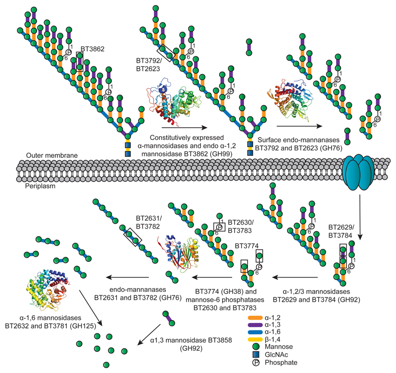
Fig. 3. Model of YM deconstruction by Bt.
Boxes show examples of bonds cleaved by the endo-α1,2-mannosidase BT3862, by the α-1,3- and α-1,2-mannosidase activities displayed by BT2629 and BT3784, and the Man-1-P and α-1,2-Man linkages targeted by BT3774. Structures of the enzymes that play a key role in mannan degradation, colour-ramped from the N (blue) to the C (red) terminus; ED Fig. 3 provides. In this model limited degradation occurs at the surface and the bulk of glycan degradation occurs in the periplasm. SusC-like proteins mediate transport across the outer membrane7.

