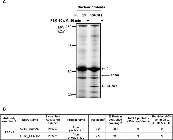
Fig 5. Nuclear association of RACK1 with β-actin.
SH-SY5Y cells were treated with 10 μM FSK for 30 min, nuclear proteins were isolated and nuclear RACK1 was immunoprecipitated. Proteins were resolved by SDS-PAGE and the gel was stained with Deep Purple™ to visualize proteins. The gel slice indicated by an arrow was in-gel protein digested and resulting peptides were submitted to mass spectrometry (MS/MS) sequencing for protein identification. B Table shows the data leading to the identification of β-actin (ACTB) and γ-actin (ACTG) proteins. Numbers refer to the legend of Fig 1B.

