
Fig 3. Analysis of the polarized cell division process of C. trachomatis serovar L2 in live and fixed cells.
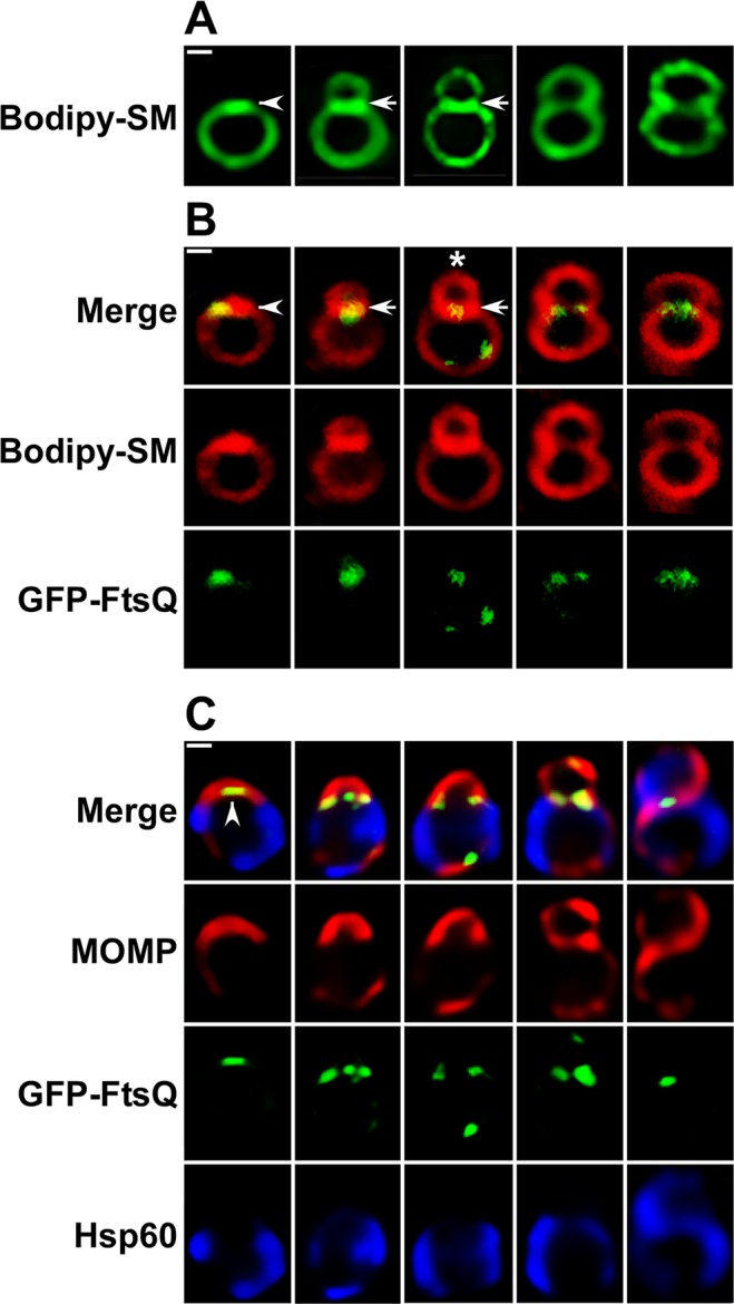
(A) HeLa cells infected with C. trachomatis were incubated in the presence of green fluorescent BODIPY FL C5 ceramide as described in the Materials and Methods and polarized cell division intermediates were imaged in live cells at 11 hours post-infection using a Zeiss AxioImager.M2 microscope. (B) HeLa cells were infected with C. trachomatis serovar L2 that contained an anhydrotetracycline (aTc)-inducible plasmid expressing GFP-FtsQ [17]. aTc was added to infected cultures at 8 hours post-infection then the cells were labeled with red fluorescent BODIPY TR C5 ceramide. Polarized cell division intermediates were imaged in live cells at 11 hours post-infection using a Zeiss LSM710 confocal microscope. Arrowheads in A and B indicate regions of intense BODIPY-sphingomyelin fluorescence at one pole of a round cell prior to division. The polar patch of sphingomyelin fluorescence in B also contained GFP-FtsQ. Arrows in A and B indicate a sphingomyelin-rich membrane separating a nascent daughter cell from a mother cell. (C) HeLa cells were infected with C. trachomatis serovar L2 that contained aTc-inducible version of GFP-FtsQ. The fusion was induced by the addition of aTc to cultures at 8 hours post-infection and the cells were fixed at 11 hours post-infection. Following permeabilization, the cells were incubated with rabbit polyclonal antibodies against Hsp60 (blue), goat polyclonal antibodies against MOMP (red), and mouse monoclonal antibodies against GFP (green) followed by donkey anti-rabbit IgG conjugated to Alexa Fluor 633, donkey anti-goat IgG conjugated to Alexa Fluor 594, and donkey anti-mouse IgG conjugated to Alexa Fluor 488 secondary antibodies. The cells were then washed prior to imaging on a Zeiss AxioImager.M2 microscope. Note the size and morphology of (B) live and (C) fixed cells are very similar. Arrowhead in C indicates a region of GFP-FtsQ fluorescence in the MOMP-positive pole of the cell prior to cell division.