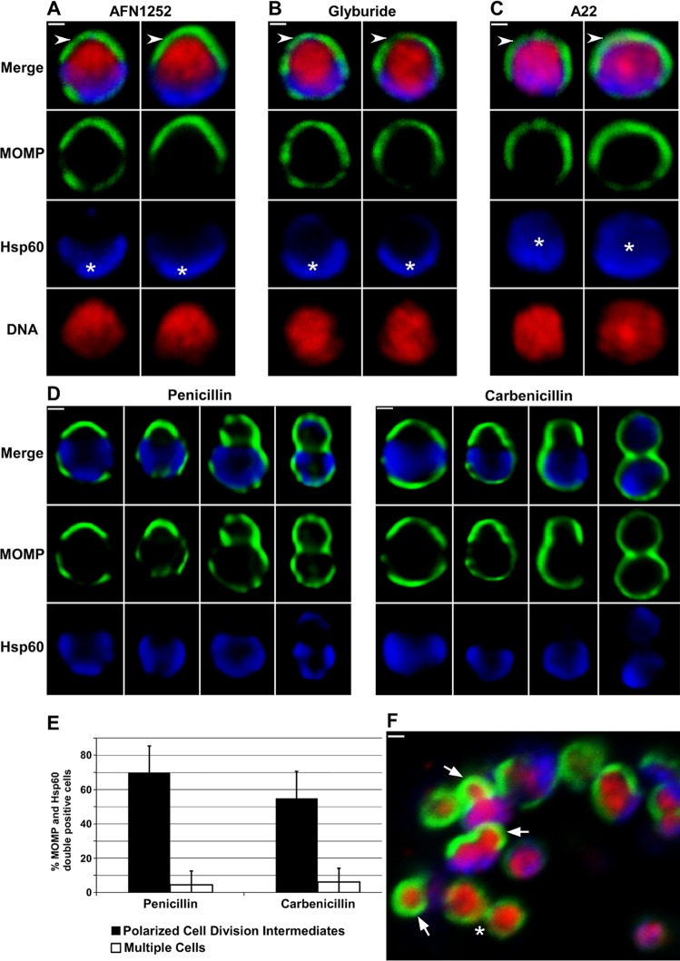
Fig 7. Inhibitors of membrane biosynthesis, peptidoglycan biosynthesis, and MreB prevent the polarized cell division of Chlamydia.
HeLa cells were infected with C. trachomatis serovar L2. At 11 hours post-infection, AFN1252 (A), glyburide (B), or A22 (C) was added to the cells and the cells were subsequently fixed at 16 hours post-infection. (D) Penicillin or carbenicillin was added to infected HeLa cells at 10 hours post-infection and the cells were fixed at 12 hours post-infection, or (F) carbenicillin was added to infected HeLa cells at 18 hours post-infection and the cells were fixed 40 minutes later. In each instance, the cells were permeabilized and incubated with rabbit antibodies against Hsp60 (blue) and goat antibodies against MOMP (green) followed by donkey anti rabbit IgG conjugated to Alexa Fluor 633 and donkey anti-goat IgG conjugated to Alexa Fluor 488. In some instances, the cells were stained with Hoechst 33342 (red) prior to microscopic analysis (A—C and F). Two cells illustrating the effect of AFN1252 (A), glyburide (B) and A22 (C) on the polarized cell division process are shown. The cells in A—C are representative of 100 cells that were analyzed from two independent experiments. Panel D illustrates the various intermediates in polarized cell division observed in penicillin-treated or carbenicillin-treated cells. Asterisks indicate polar (A and B) and diffuse (C) Hsp60. Arrows in F point to polarized cell division intermediates observed following carbenicillin treatment of a more mature inclusion containing multiple cells. The asterisk in F indicates a cell that has almost completed division. Images in A-C and F were acquired by confocal microscopy; images in D were acquired by epifluorescent microscopy. White bars are 0.5μ. (E) Penicillin or carbenicillin were added to infected HeLa cells at 10 hours post-infection and the cells were fixed at 12 hours post-infection. The percentage of C. trachomatis that were undergoing polarized cell division (black bars) or had completed the first division (white bars) were quantified. All of the MOMP and Hsp60 double-positive cells in randomly selected fields were included in the analysis. The data shown represents the analysis of more than 200 cells from two independent experiments.

