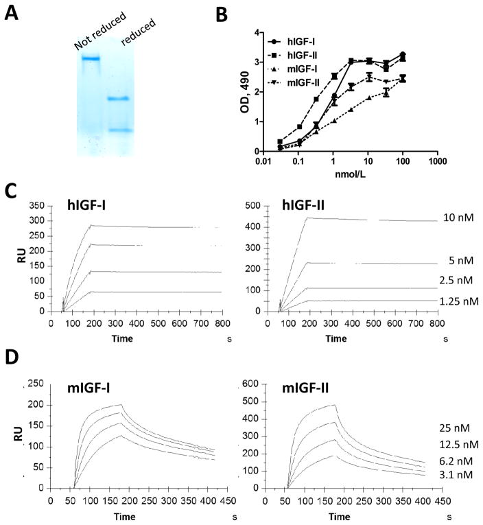
Figure 1. Characterization of IgG1 m708.5.
A, The purity of purified IgG1 m708.5 was analyzed by SDS-PAGE under native and denaturing conditions. B, Binding of IgG1 m708.5 to hIGF-1, huIGF-2 and mIGF-1, mIGF-2 was analyzed by ELISA. IgG1 m708.5 were serially diluted and added to wells coated with IGF-1 or IGF-2. Bound IgG1 was detected with an HRP conjugated anti-human Fc antibody and optical densities (O.D.) measured at 490 nm. C and D, sensorgrams of binding kinetics of IgG1 m708.5 as measured with Biacore. The hIGF-1, hIGF-2 (C) or mIGF-1, mIGF-2 (D) were immobilized and different concentrations of the antibodies were used as indicated in the figures. Details of the experiments were described in the Materials and Methods.

