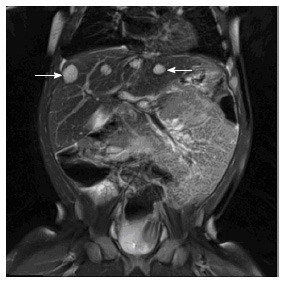
Figure 2.

Multiple infantile hepatic hemangiomas. Coronal T2 weighted MRI image through the upper abdomen in a 5-mo-old girl depicts multiple well-defined, T2 hyperintense masses in the liver (arrows). This was consistent with multifocal infantile hepatic hemangiomas. MRI: Magnetic resonance imaging.
