Figure 3.
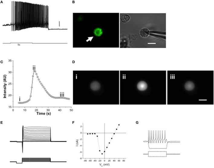
Melanopsin expression confers intrinsic photosensitivity to TG neurons. (A) 5 s 480 nm stimulus initiated delayed onset firing of action potentials in current clamp recordings. Scale bar = 10 mV. (B) Dissociated OPN4 trigeminal neurons were identified by EGFP fluorescence (left panel, arrow). Trigeminal neurons not expressing EGFP were identified by brightfield illumination (right panel, arrowhead). Scale bar = 20 μm. (C) Constant 480 nm light stimulated slow, inactivating calcium signals in some isolated TG neurons from wild type mice, measured with fluo-4 AM. (D) Freeze frame [Ca2+]i images taken before stimulation (i), at the peak of the Ca2+ signal (ii) and after return to normal (iii). Scale bar = 20 μm. (E) Voltage clamp recording showing Na+ and K+ currents in response to voltage clamp steps of 100 ms duration. (F) Current-voltage relation of isolated TG neuron shown in E. (G) Current clamp recording of an isolated TG neuron showing response to 10 pA hyperpolarizing and depolarizing current injections, the latter producing a train of action potentials. The current injection steps lasted 100 ms.

