Figure 6.
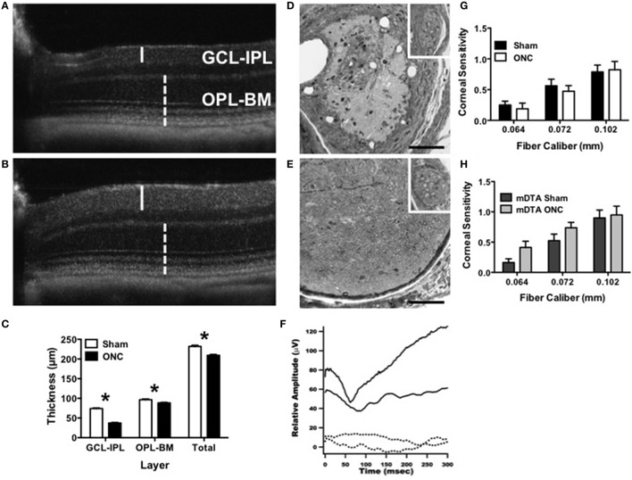
ONC causes degeneration of RGCs and the optic nerve with no alteration to corneal innervation. (A–C) Degeneration of the ganglion cell layer 45 days after ONC was assessed using in vivo sd-OCT imaging. Representative images are shown for (A) ONC and (B) sham. The inner retina containing retinal ganglion cells (GCL-IPL, solid line) showed greater degeneration than the outer retina containing rod and cone photoreceptors (OPL-BM, dashed line). (C) The average thickness for sham and ONC is shown for the GCL-IPL (T14 = 21.4, p < 0.0001), OPL-BM (T14 = 3.6, p = 0.003) and the total retinal thickness (Total, T14 = 6.1, p < 0.0001). (D,E) Optic nerve damage was assessed by light microscopy with representative cross-sectional images approximately 0.5 mm from the globe shown for (D) ONC and (E) sham. Inset shows intact trigeminal nerve bundles. Scale bar = 100 μm. (F) OPN4dta/dta mice with sham (solid line) showed normal P1-N1 response amplitudes whereas the VEP was not recordable in OPN4dta/dta mice with ONC (dashed line). (G,H) Trigeminal innervation of corneal mechanosensitivity was tested using von Frey fibers (0.064–0.102 mm diameter) in (G) OPN4+/+ mice with sham (black, n = 16 eyes) or ONC (white, n = 16 eyes) and (H) OPN4dta/dta mice with sham (black, n = 16 eyes) or ONC (gray, n = 16 eyes), showing the same corneal sensitivity (no interaction of surgery by fiber caliber with no effect of surgery but a main effect of fiber caliber [F(2, 90) = 17, p < 0.0001, F(2, 78) = 14, p < 0.0001, respectively]). Data are represented as mean ± SEM, students t-test and p-values are given. * p < 0.05.

