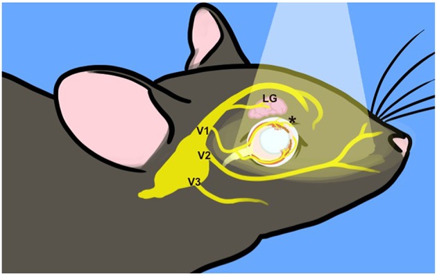
Figure 7.

Incident light reaches trigeminal nerve terminals. Surface structures innervated by the trigeminal nerve are exposed to incident light, indicated by the pale cone. The TG are situated at the base of the skull. Three major branches exit the skull to target ocular (V1, ophthalmic), upper jaw (V2, mandibular), and lower jaw (maxillary) structures. The frontal branch of V1 innervates the lacrimal gland (LG) and forehead. The ciliary branch innervates the cornea, iris and choroid (*) as well as the nasal mucosa (not depicted). For illustration purposes, many trigeminal branches are not shown and the depiction is not to scale.
