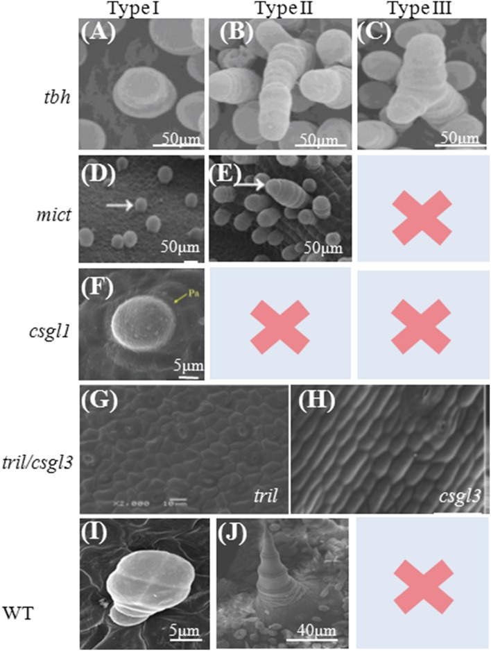
Figure 2.
Scanning Electron Microscopy images of trichomes of cucumber mutants. (A–C) SEM images of fruit spines (A,B) and leaf trichomes (C) in tbh, three morphologies from left to right; (D–E). Micro-trichomes on a mict leaf. Arrows indicate the two morphologies: type I (right) and type II (left), respectively; (F). Trichomes on the epidermis of leaves from csgl1; (G,H). The tril/csgl3 mutant has a completely glabrous phenotype, without trichome material in fruit epidermis. Trichome on the Fruit from the Wild type as control (I,J). Pictures (A–C) are from (Chen et al., 2014); Pictures (D,E,H) are from (Zhao et al., 2015a); Picture (F) is from (Li et al., 2015), and Picture (G) is from (Wang et al., 2016).

