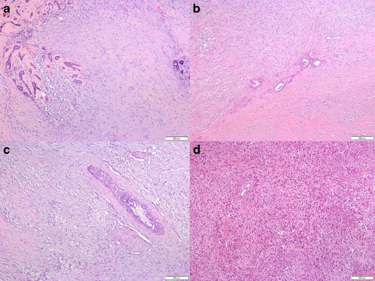
Fig. 2.
Spindle cell myoepithelial carcinomas (SCMCs) were characterized by proliferation of spindle cells with slender nuclei in varying densities, infiltrating around pre-existing ducts and lobuli (a–c). All tumors exhibited matrix production but to varying extents (a, b). Due to mucoid change of intercellular matrix, pseudoangiomatoid spaces occurred in some cases (a, lower left quadrant) with an endothelial-like inner cell layer around the mucoid material. Occasionally, foci of squamous differentiation not exceeding 10 % of tumor cells were found (c). Brain metastasis retained spindle cell morphology but revealed a higher cell density (d) (a–d, hematoxylin-eosin stain)

