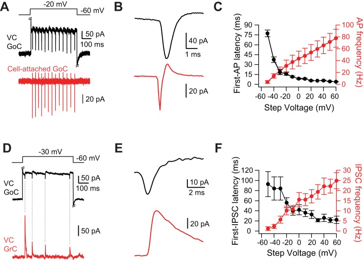
Fig. 4.
Depolarization-triggered spikelets originate in Golgi cells. A: step to −20 mV from a holding potential of −60 mV in the voltage-clamped Golgi cell evoked fast inward currents (spikelets) in the voltage-clamped Golgi cell (black trace) and spikes in the Golgi cell (red) simultaneously recorded in the cell-attached configuration. Fast chemical synaptic transmission blocked by strychnine, SR 95531, NBQX, and R-CPP. Intracellular solution for black Golgi cell was CsCl based and contained QX-314. B: spike-triggered average of spikelets from same pair as in A. C: latency to first extracellularly recorded AP from the beginning of the voltage step (black) and frequency of extracellularly recorded APs (red) as a function of the voltage step (n = 4). D: step to −20 mV from a holding potential of −60 mV evoked spikelets in the Golgi cell (black) and IPSCs onto the simultaneously recorded voltage-clamped granule cell (red). Excitatory synaptic transmission blocked by NBQX and R-CPP. Intracellular solution for Golgi cell was CsCl based and contained QX-314. E: IPSC-triggered average of spikelets from same pair as in D. Note that depolarizing junctional current precedes IPSC. F: latency to first IPSC onto the granule cell from the beginning of the voltage step (black) and frequency of IPSCs onto granule cells (red) as a function of the voltage step (n = 18). Error bars show SE.

