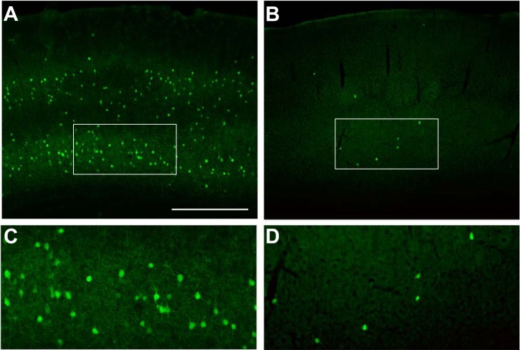
Fig. 1.
Putative PV+ interneurons (GFP+) are abundant and broadly distributed in the G42 mouse cortex (A). Imaging layer 5/6 under higher magnification (white box) reveals the density and morphology of these mature interneurons (C). Small, single-site transplantation of MGE progenitors from G42 mouse embryos results in a broad but sparse distribution of GFP+ cells (B). Shown is a section ∼300 μm anterior to the injection site. Imaging at higher magnification reveals mature neuronal morphology similar (D) to the cells in their native environment. Scale bar in A represents 500 μm for A and B and 200 μm for C and D.

