Fig. 4.
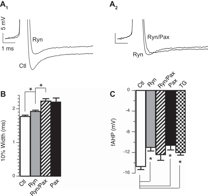
Ca2+ release through ryanodine receptors (RyR) activates BK during the fAHP. A1: representative threshold APs in the absence or presence of 20 μM ryanodine (Ryn) in β4KO neurons. A2: representative threshold APs in the absence or presence of 5 μM Pax in addition to 20 μM Ryn. B: averaged 10% width in ms: control 1.77 ± 0.04 (n = 24); Pax 2.20 ± 0.12 (n = 8); Ryn 1.92 ± 0.04 (n = 13); Ryn/Pax 2.23 ± 0.07 (n = 9); Ryn and control are significantly different (P ≈ 0.03). Ryn/Pax and Pax are not significantly different (P = 0.82). C: Ryn significantly decreased fAHP amplitudes. Averaged fAHP amplitudes in mV: control −14.7 ± 0.6 (n = 24); Pax −10.6 ± 0.9 (n = 8); Ryn −11.0 ± 0.7 (n = 13); Ryn/Pax −12.4 ± 1.1 (n = 9); thapsigargin (TG) −12.0 ± 0.5 (n = 6). Ryn and control are significantly different (P = 0.0005). Ryn/Pax and Ryn are not significantly different (P = 0.30). Ryn/Pax and Pax are not significantly different (P ≈ 0.24). TG (1 μM) and control are significantly different (P ≈ 0.03). TG and Ryn are not significantly different (P ≈ 0.41). *P < 0.05.

