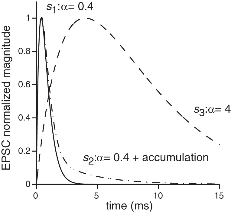
Fig. 2.

Three EPSC shapes were used to examine the impact of EPSC shape on spike response patterns. The first shape, s1(t), was derived by fitting an alpha function to the synaptic currents recorded from the terminals contacting inner hair cells (e.g., Glowatzki and Fuchs 2002; Grant et al. 2010); these shapes are similar to most events from calyx terminals (Sadeghi et al. 2014). The second shape, s2(t), had a fast onset time [same as s1(t)] but an elongated decay. Such shapes could result from glutamate accumulation and other complexities related to the unique synaptic cleft of calyx terminals. The third shape, s3(t), had a long onset and decay phase that could result from a variety of factors, including the slowing and attenuation of synaptic events via dendritic filtering. Detailed description and equations are provided in the text.
