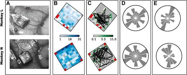
Figure 6.
Spatial arrangement of neurons participating in significant patterns. A, Locations of the electrode arrays in the motor cortex in the right hemisphere of the two monkeys. CS, Central sulcus; AS, arcuate sulcus; PD, precentral dimple. B, Color maps of the total number of SUAs recorded on each electrode of the recording array summed across all selected sessions. Red squares indicate the four unconnected electrodes. C, Color maps showing, for each electrode, the total number of patterns found that involved neurons recorded on that electrode. The number is obtained separately for each session (summing across all epochs and trial types) divided by the total number of single neurons recorded on the electrode in the session and finally summed across sessions. The final value can exceed the number of sessions (10) if, for instance, in each session there were more significant patterns involving the electrode than neurons recorded by that electrode. Gray squares correspond to a value of 0 and indicate electrodes that never recorded single neurons involved in significant patterns. Black lines connect electrode pairs extracted from each significant pattern found. D, Number of all possible pairs of recorded neurons placed along each orientation. Each panel shows a bar chart in polar coordinates. The direction of each bar corresponds to one orientation on the recording array, whereas the length of the bar represents the total number of neuron pairs placed along that direction computed for each session and summed across sessions. The circles have a diameter of 5000 for Monkey L and of 18,000 for Monkey N. E, Spatial orientation of all pairs of neurons involved in significant patterns found during movement as a fraction over the total number of recorded neuron pairs with that orientation shown in D.

