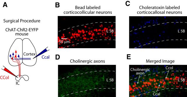
Figure 3.
ChR2-EYFP fibers among AC L5B corticocallosal and corticocollicular neurons in the ChAT-ChR2-EYFP mouse line. A, Labeling of corticocollicular and corticocallosal neurons with fluorescent tracers. Projection neurons in the AC were labeled by injecting different colored retrograde tracers in the contralateral AC (choleratoxin to label corticocallosal neurons) and the ipsilateral IC (red fluorescent microspheres to label corticocollicular neurons). B, A 20× epifluorescence image showing labeled corticocollicular neurons in L5B of the AC. C, A 20× epifluorescence image showing labeled corticocallosal neurons in L5B and other layers of the AC. D, A 20× epifluorescence image showing green cholinergic axons in L5B of AC. E. Merged image (B–D combined) showing intermingled population of corticocallosal and corticocollicular neurons among green cholinergic axons in L5B of AC.

