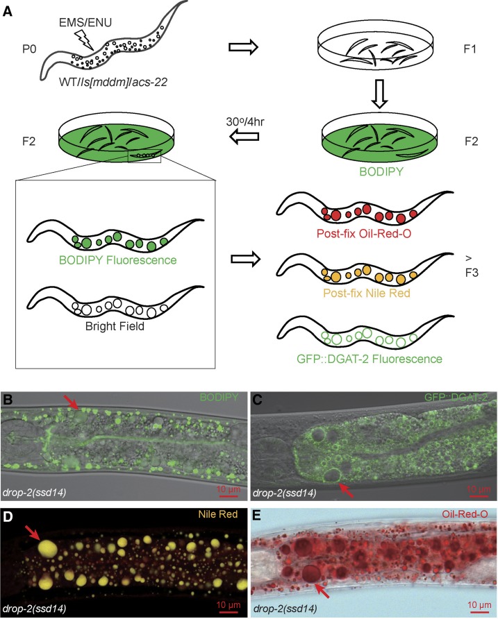
Figure 1.
The design of mutagenesis and LD labeling. (A) Both EMS and ENU were used to mutagenize wild type (WT), transgenic lines ssdIs1 and ssdIs5[maoc-1; dhs-28; daf-22; mrfp::pts1] (Is[mddm]), and acs-22(tm3236) P0 animals. F2 progeny were synchronized and transferred onto NGM/OP50 plates layered with BODIPY and were allowed to grow to adulthood at 15° or 20° before being shifted to 30° for 4 hr. Candidate mutants were isolated by virtue of BODIPY-positive and light-refracting globular structures visible under a fluorescence stereoscope at 180× magnification. F3s and further generations of F2 isolates were then tested by postfix Oil-Red-O staining, postfix Nile Red staining, and were crossed into hjSi56[gfp::dgat-2] for testing GFP::DGAT-2 marking. Supersized LDs (arrows) in drop-2(ssd14), one of the 118 isolates, were readily visible in bright field and were labeled by vital BODIPY (B), by GFP::DGAT-2 (C), by postfix Nile Red with a true color of gold (D), and by postfix Oil-Red-O (E). (B) and (C) are channel mode confocal microscopy images; (D) lambda mode confocal microscopy, also note the diffuse red fluorescence of membrane structures labeled by postfix Nile Red; (E) wide field microscopy.

