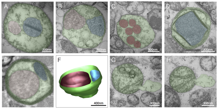
Figure 5. Compartmentalization.
(A) Example of membrane-bound sub-mitochondrial compartments located centrally and (B) peripherally in contact with the inner boundary membrane, from cases of m.8344A>G mutation (patient 5) and single mtDNA deletion (patient 3), respectively. (C) Electron-dense round compartments distributed in the mitochondrial matrix in a case of single mtDNA deletion (patient 3). (D) Compartment bound by linearized electron-dense membranes in a case of m.8344A>G (patient 4). (E) Cross-sectional image of a mitochondrion with two distinct compartments of different electron density, and (F) three-dimensional reconstruction from SBF-SEM. (G,H) Examples of OMM protrusion and distension consistent with the release of mitochondrial components in the cytoplasm in a case of m.8344A>G mutation (patient 4). Pseudocolored areas indicate the major compartment bound by the OMM (green), and sub-compartments (red and blue).

