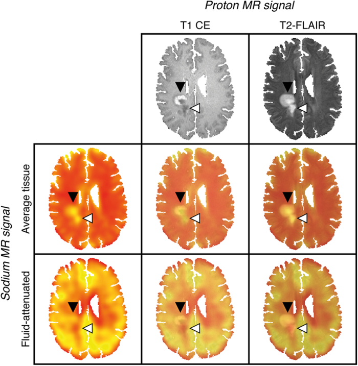
Figure 1. Exemplary co-registered sodium and proton images of a patient with acute MS lesions (PID no. 15).

Proton MRI demonstrates two (confluent) right-central white matter lesions. The anterior lesion exhibits contrast-enhancement and corresponding elevated average tissue and fluid-attenuated sodium signals compatible with acute inflammation (black arrowhead). The posterior lesion shows no signs of blood-brain barrier disruption, an increased average tissue sodium signal and a reduced fluid-attenuated sodium signal – a combination consistent with the residuals of brain tissue inflammation (white arrowhead). This representative example demonstrates high intermodal registration accuracy of the applied affine image transformation method (FLIRT); T1 CE, T2-FLAIR and sodium images as well as the corresponding overlays are shown.
