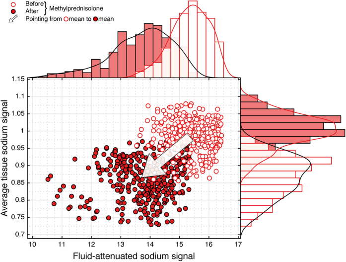
Figure 3. Longitudinal sodium MRI data of an acute lesion (PID no. 15).
After application of high-dose methylprednisolone both the average tissue and fluid-attenuated sodium signal decrease. Based upon the cross sectional findings on sodium signal differences between acute and chronic MS lesions, this signal behavior was proposed before. Moreover, it strongly supports the notion that findings of our study are compatible with intracellular sodium accumulation in acute inflammatory MS lesions. Please note, that due to the pharmacological intervention changes in tissue sodium concentration of healthy parenchyma could not be ruled out. Thus, for the longitudinal observations, normalization referred to transmitter amplitudes instead of the sodium signal of healthy parenchyma.

