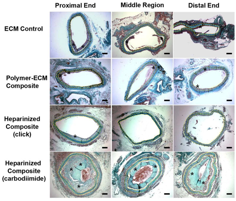
Figure 6.
Masson’s Trichrome stain of ECM control, polymer-ECM composite, heparinized polymer-ECM composite prepared via “click” chemistry, and heparinized polymer-ECM composite prepared via carbodiimide chemistry at the proximal end, middle region and distal end of the graft. All vascular grafts were recovered after implantation as an aortic interposition graft at 4 weeks after surgery and all microscopic images were taken with a 4x objective. Grafts are outlined between yellow dashed lines, while * indicates the presence of intimal hyperplasia. Scale bar = 200 μm.

