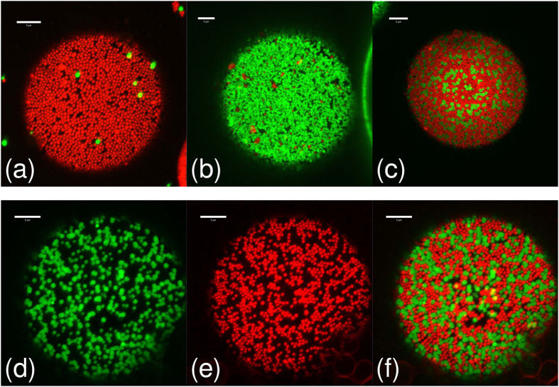
Figure 4. Droplet interface details.
High magnification confocal micrographs of the ΦIM = 50% sample from Fig. 3(a–e) showing the particle monolayers at droplet interfaces at various rolling times. The rolling times are (a) 0 h, (b) 5.5 h, (c) 22 h and (d–f) 357 h. Images (d–f) are of the same droplet, showing (d) the green channel, (e) the red channel and (f) the merged image. Each image is a z-projection of 10 images, with a z-spacing of 0.42 μm, constructed using ImageJ28. All scale bars are 5 μm.

