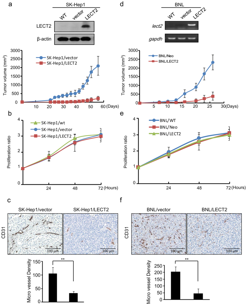
Figure 1. Ectopic LECT2 expression inhibits tumor growth and angiogenesis in an HCC xenograft model.
(a) Top, analysis of stable expression of LECT2 protein in SK-Hep1 cells by immunoblotting. Bottom, tumor volume was measured by using a two-dimensional caliper at regular intervals in NSG mice inoculated subcutaneously with control or LECT2-expressing SK-Hep1 cells. (b) The proliferation ratios of SK-Hep1 cells as determined using an MTT assay for 3 days. Each data point is representative of three independent experiments and presented as the mean ± SD. (c) The effects of LECT2 expression on tumor angiogenesis and growth in a xenograft mouse model of HCC. Top, sections of tumors obtained from mice were stained with the specific murine blood vessel marker CD31. Bottom, quantitation of MVD in the xenograft tumors obtained from mice. (d) Top, analysis of lect2 gene expression in stable BNL cells by reverse transcription-polymerase chain reaction. Bottom, tumor volume was measured by using a two-dimensional caliper at regular intervals in BALB/C mice inoculated subcutaneously with control or lect2-expressing BNL cells. (e) The proliferation ratios of BNL cells as determined using an MTT assay for 3 days. (f) The effects of lect2 expression on tumor angiogenesis and growth in a xenograft mouse model of HCC. Top, sections of tumors obtained from mice were stained with CD31. Bottom, quantitation of MVD in the xenograft tumors obtained from mice.

