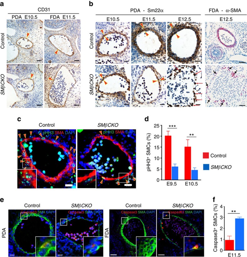
Figure 2. β-Catenin promotes SMC proliferation and survival during artery formation.
(a) Immunohistochemistry (IHC) for the endothelial marker CD31. Arrowheads indicate the endothelial layer. Scale bar, 50 μm. (b) IHC for SMC markers, Sm22α (brown; scale bar, 25 μm) and α-SMA (red; scale bar, 50 μm). Arrowheads delimit the vessel wall. Arrows indicate scattered SMCs. (c) Immunostaining of PDAs for the mitotic marker pHH3 and α-SMA at E10.5. Arrowheads indicate pHH3+ SMCs. Scale bar, 20 μm. (d) Quantification of pHH3+ SMCs in the wall of PDAs. **P<0.01; ***P<0.001 comparing genotypes by two-way ANOVA and Sidak's multiple comparison test. n=6. (e) Immunostaining for cleaved Caspase3 and α-SMA at E11.5. Scale bar, 20 μm (PDA); 50 μm (FDA). (f) Quantification of Caspase3+ SMCs in the arterial wall. **P<0.01 comparing genotypes by two-tailed t-test. n=3. In d,f, data represent the mean±s.e.m.

