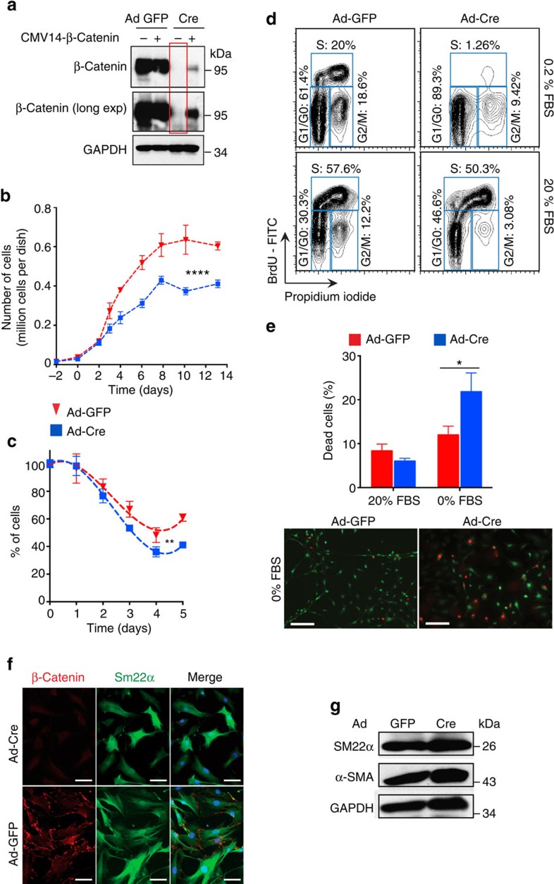
Figure 3. β-Catenin is required for vascular SMC population growth.
(a) Western blot analysis of indicated proteins in mouse aortic SMCs isolated from Ctnnb1flox/flox mice, transduced with GFP- or Cre-expressing adenovirus (Ad-GFP or Ad-Cre) and transfected with CMV14-β-catenin or empty vector. β-Catenin expression is not detectable in Ad-Cre SMCs even with a long exposure (red box). β-Catenin expression restored by lipid-based transfection of CMV14-β-catenin in Ad-Cre SMCs (fourth lane). (b) Mouse aortic SMC population growth under standard growth conditions. ****P<0.0001 comparing Ad-GFP versus Ad-Cre by two-way ANOVA. (c) Mouse aortic SMC population decline under serum starvation. **P<0.01 comparing Ad-GFP versus Ad-Cre by two-way ANOVA. (d) FACS analysis of BrdU and propidium iodide staining in mouse aortic SMCs with the indicated treatments and in both low and high serum conditions. (e) Top: quantification of dead cells. *P<0.05 comparing Ad-GFP versus Ad-Cre by two-way ANOVA and Sidak's multiple comparisons test. Bottom: LIVE/DEAD assay using mouse aortic SMCs with the indicated treatments. Live cells in green; dead cells in red. Scale bar, 200 μm. (f) Immunocytochemistry, scale bar, 50 μm and (g) western blot analysis of β-catenin and indicated SMC markers in Ad-GFP or Ad-Cre-transduced mouse aortic SMCs. In b,c,e, data represent the mean±s.e.m. and n=3.

