Figure 1.
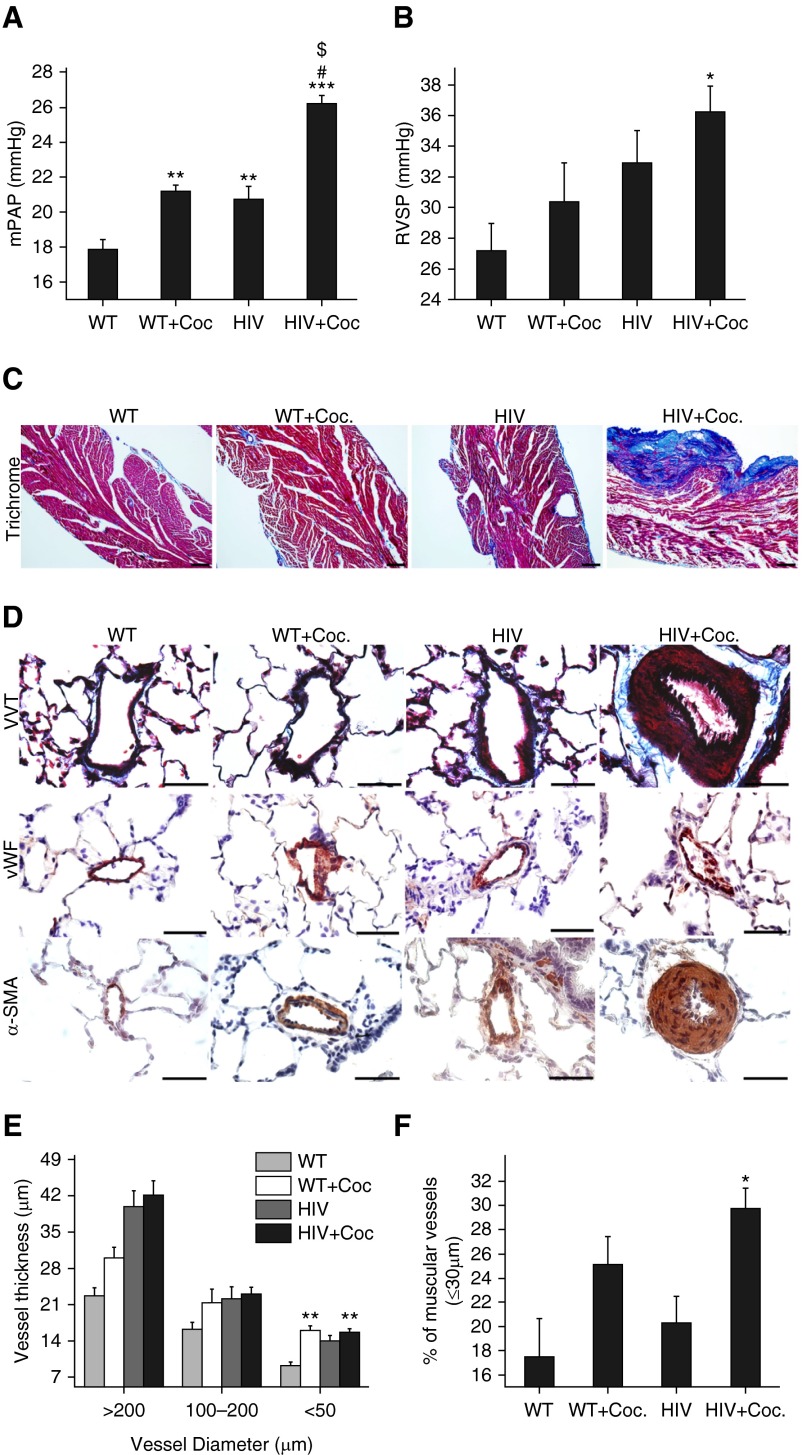
Hemodynamics and pulmonary arterial remodeling in 4-month-old human immunodeficiency virus (HIV)–transgenic (Tg) rats exposed to cocaine. The 4-month-old male Fischer HIV-Tg rats were administered 40 mg/kg body weight of cocaine (HIV + Coc. group) or saline (HIV group) daily for 21 days. Wild-type (WT) F334 rats with (WT + Coc. group) and without cocaine (WT group) were used for comparison. (A) Mean pulmonary arterial pressure (mPAP) and (B) right ventricle (RV) systolic pressure (RVSP) were measured after RV catheterization. Values are mean (±SEM) of n ≥ 6 per group. (C) Masson’s trichrome staining in snap-frozen, formaldehyde-fixed, paraffin-embedded heart sections. Scale bars: 100 μm. (D) Lungs were harvested, fixed, paraffin-embedded, and sectioned followed by staining for Verhoeff-Van trichrome (VVT), von Willebrand factor (vWF), and α-smooth muscle actin (α-SMA). Scale bars: 50 μm. (E) Thickening of vessels was assessed by measuring inner and outer diameter of various-sized vessels. Significant increase in the thickness of arteries with a diameter less than 50 μm was observed in the HIV + Coc. group. (F) α-SMA–positive vessels with an outer diameter less than 30 μm were counted per rat within each group (n = 5 rats/group) to determine the percentage of muscularization of intra-acinar vessels. *P < 0.05, **P < 0.01, and ***P < 0.001 versus WT, #P < 0.001 versus WT + Coc., $P < 0.001 versus HIV.

