Figure 1.
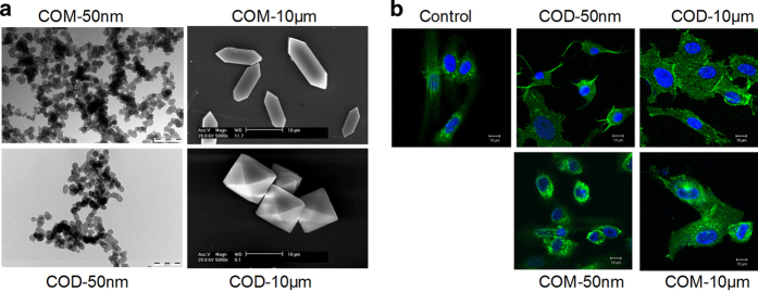
Morphological observation of nano-/micron-sized COM and COD crystals and cytoskeleton. (a) SEM and TEM images of nano-/micron-sized COM and COD crystals, respectively. (b) Confocal laser scanning microscopy observation of the changes of cytoskeleton caused by nano-/micron-sized COM and COD crystals. Cell nuclei (blue) and cytoskeleton (green, as represented by F-actin) were stained with DAPI and FITC-conjugated phalloidin, respectively. Crystal concentration: 200 μg/ml.

