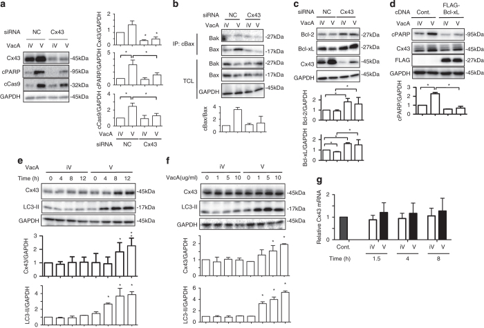
Figure 1.
Increased Cx43 protein in VacA-treated AZ521 cells. (a) Control (NC) and Cx43 siRNA-transfected AZ-521 cells were incubated with 120 nM heat-inactivated (iV) or wild-type VacA (V) for 8 h, lysed with 1× SDS sample buffer and analyzed by immunoblotting with the indicated antibodies. Quantification of VacA-induced Cx43, cPARP and cCas9 levels in AZ-521 cells was performed by densitometry (right panel). Data are presented as mean±S.D. of values from three experiments and significance is *P<0.05. Experiments were repeated three times with similar results. (b) The indicated siRNA-transfected cells were treated with 120 nM heat-inactivated (iV) or wild-type VacA (V) for 6 h, lysed with cell lysis buffer containing 2% CHAPS and immunoprecipitated with anti-conformational changed Bax-specific antibody (cBax) as described in Materials and Methods. Experiments were repeated three times with similar results. Quantification of VacA-induced cBax levels in AZ-521 cells was performed by densitometry (bottom panel). (c) The indicated siRNA-transfected AZ-521 cells were incubated with 120 nM heat-inactivated (iV) or wild-type VacA (V) for 8–10 h, lysed with cell lysis buffer containing 1% NP40 and analyzed by immunoblotting with the indicated antibodies. Quantification of VacA-induced Bcl-2 and Bcl-xL levels in AZ-521 cells was performed by densitometry (bottom panels). Data are presented as mean±S.D. of values from four experiments and significance is *P<0.05. Experiments were repeated three times with similar results. (d) Control (Cont.) or FLAG-tagged Bcl-xL plasmid-transfected cells incubated with 120 nM heat-inactivated (iV) or wild-type VacA (V) for 10 h, lysed with 1× SDS sample buffer and analyzed by immunoblotting with the indicated antibodies. Quantification of VacA-induced cPARP levels in AZ-521 cells was performed by densitometry (bottom panel). Data are presented as mean±S.D. of values from three experiments and significance is *P<0.05. Experiments were repeated three times with similar results. (e) AZ-521 cells were incubated with 120 nM heat-inactivated (iV) or wild-type VacA (V) for the indicated times, lysed with 1× SDS sample buffer and analyzed by immunoblotting with the indicated antibodies. Quantification of VacA-induced Cx43 levels in AZ-521 cells was performed by densitometry (bottom panel). Data are presented as mean±S.D. of values from three experiments and significance is *P<0.05. Experiments were repeated three times with similar results. (f) AZ-521 cells were incubated with the indicated concentration of heat-inactivated (iV) or wild-type VacA (V) for 10 h, lysed with 1× SDS sample buffer and analyzed by immunoblotting with the indicated antibodies. Quantification of VacA-induced Cx43 levels in AZ-521 cells was performed by densitometry (bottom panel). Data are presented as mean±S.D. of values from three experiments and significance is *P<0.05. Experiments were repeated three times with similar results. (g) AZ-521 cells were incubated with 120 nM heat-inactivated (iV) or wild-type VacA (V) for the indicated times and total RNA was extracted as described in Materials and Methods. Cx43 mRNA was measured by real-time qPCR. Data are shown as mean±S.D. of values from three experiments. Results are shown as fold increase of GAPDH as an internal control. Experiments were repeated three times with similar results.

