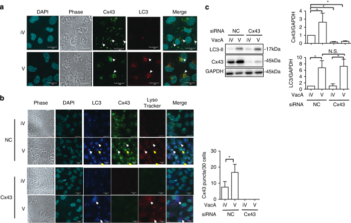
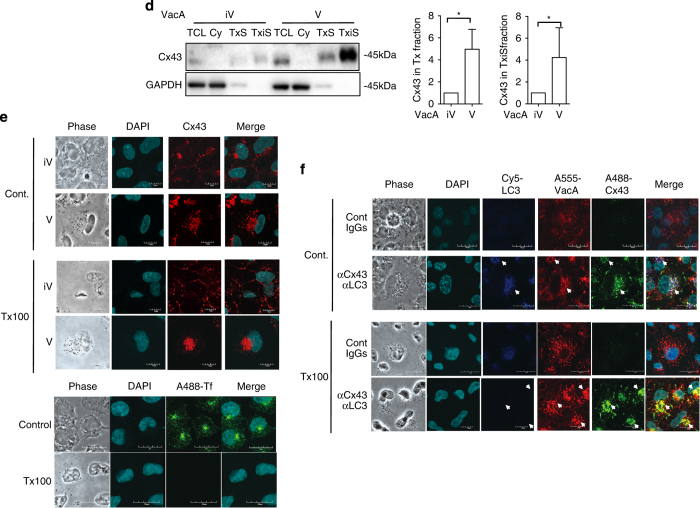
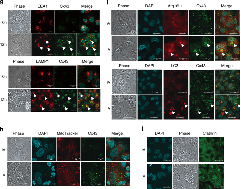
Figure 3.
VacA-increased Cx43 colocalized with EEA1, LAMP1, Atg16L1 and LC3 in Tx-insoluble compartment. (a) AZ-521 cells were incubated with 120 nM heat-inactivated (iV) or wild-type VacA (V) for 10 h, then reacted with anti-Cx43 antibodies (green) and with DAPI (cyan). The arrows indicate Cx43 in plasma membranes. Bars represent 20 μm. Experiments were repeated three times with similar results. (b) AZ-521 cells were incubated with 120 nM heat-inactivated (iV) or wild-type VacA (V) for 10 h and 100 nM LysoTracker (red) added to cells before fixation as described in Materials and Methods. Cells were reacted with anti-Cx43 antibodies (green) and DAPI (cyan). Merged and higher magnification images of the outlined areas are shown. Bars represent 20 μm. Experiments were repeated two times with similar results. The arrows indicate the colocalization of LC3, Cx43 and LysoTracker. The number of Cx43 puncta in a single cell was manually counted under a confocal microscopy (right panel; *P<0.05). For each group, 30 cells were randomly selected for the average of number of Cx43 puncta in the cell. (c) The indicated siRNA-transfected cells were incubated with 120 nM heat-inactivated (iV) or wild-type VacA (V) for 10 h. Cells were lysed with 1× SDS sample buffer and analyzed by immunoblotting with the indicated antibodies. Quantification of VacA-induced Cx43 and LC3-II generation in AZ-521 cells was performed by densitometry (right panel). Data are presented as mean±S.D. of values from three experiments and significance is *P<0.05. Experiments were repeated three times with similar results. (d) After cells treated with heat-inactivated (iV) or wild-type VacA (V) for 10 h, cells were fractionated as described in Materials and Methods. TCL, total cell lysate; Cy, cytoplasm and small organelles; TxS, Triton X-100 soluble fraction. TxiS, Triton X-100 insoluble fraction. Proteins were applied to SDS-PAGE, followed by immunoblotting with the indicated antibodies. Quantification of VacA-induced Cx43 levels in Tx-soluble and TxiS-soluble fraction was performed by densitometry (bottom panel). Data are presented as mean±S.D. of values from three experiments and significance is *P<0.05. Experiments were repeated three times with similar results. (e) AZ-521 cells were incubated with 120 nM heat-inactivated (iV) or wild-type VacA (V) for 10 h and were treated with or without cold 1% Tx, fixed with 4% PFA, and reacted with anti-Cx43 antibodies (red) and incubated with DAPI (cyan) as described in Materials and Methods. Alexa488-labeled Transferrin was used as a positive control. Bars represent 20 μm. Experiments were repeated three times with similar results. (f) AZ-521 cells were incubated with Alexa555-labeled VacA for 10 h and then were treated with or without cold 1% T×100, fixed with 4% PFA, and reacted with anti-Cx43 antibodies (green), anti-LC3 antibodies (blue) and with incubated DAPI (cyan). Bars represent 20 μm. Experiments were repeated three times with similar results. The arrows indicate the colocalization of VacA, Cx43 and LC3. (g) AZ-521 cells were incubated with 120 nM heat-inactivated (iV) or wild-type VacA (V) for 10 h and then reacted with anti-EEA1 or anti-LAMP1 (red), or anti-Cx43 antibodies as indicated (green) and incubated with DAPI. Bars represent 20 μm. Experiments were repeated three times with similar results. The arrows indicate the colocalization of Cx43 and EEA1 or LAMP1. (h) AZ-521 cells were incubated with 120 nM heat-inactivated (iV) or wild-type VacA (V) for 10 h; 50 nM MitoTracker (red) was added to cells before fixation as described in Materials and Methods. Cells were reacted with anti-Cx43 antibodies (green) and incubated with DAPI (cyan). Merged and higher magnification images of the outlined areas are shown. Bars represent 20 μm. Experiments were repeated two times with similar results. (i) AZ-521 cells were incubated with 120 nM heat-inactivated (iV) or wild-type VacA (V) for 10 h, then reacted with anti-Atg16L1 antibodies (red), and anti-Cx43 antibodies (green) and incubated with DAPI. Bars represent 20 μm. Experiments were repeated three times with similar results. The arrows indicate the colocalization with Cx43 and Atg16L1. (j) AZ-521 cells were incubated with 120 nM heat-inactivated (iV) or wild-type VacA (V) for 10 h. Cells reacted with anti-clathrin antibodies (green) and incubated with DAPI (cyan). Bars represent 20 μm. Experiments were repeated two times with similar results.


